Digestion involves the chemical and physical breakdown of food into smaller molecules. This needs to happen if the molecules are to pass through the intestine wall and be transported by the bloodstream to locations in the body where they are needed.
Enzymes
Enzymes are proteins that facilitate chemical reactions. They make life processes possible by speeding up chemical reactions and lowering the temperature at which the reactions can occur. The high temperatures required by non–facilitated chemical reactions would destroy living tissue. Vitamins can be thought of as co–enzymes that assist enzymes with metabolic processes.
Enzyme–facilitated chemical reactions include such actions as molecule construction, energy transfer, and digestion. Food encounters enzymes first in the mouth, and then in numerous places along the digestive tract.
The action of salivary amylase begins carbohydrate digestion in the mouth. Amylase ("amly" is Latin for starch) is an enzyme that breaks down starch (a polysaccharide sugar) into the double sugar maltose (two linked glucose molecules). The human body cannot absorb the disaccharide sucrose, but it can absorb the simple sugars glucose and fructose.
In the classroom, the following activity can be used to simulate the action of salivary amylase. Groups of students can be asked to volunteer to chew unsweetened bread or crackers, cheese, nuts, or other similar foods for two minutes, and notice how it tastes. Ask students not to swallow (this is difficult) and then keep the bolus (chewed food) in their mouths for an additional thirty seconds. Have students make note of the new taste, then they may swallow.
The action of salivary amylase in breaking the carbohydrates into sugars in the mouth can be observed by the sweeter taste of the starch (bread or crackers) after the two minutes of chewing. Encourage students to discuss their results with each other.
As with the molecular modeling suggested for building carbohydrates, a classroom activity around disassembling the model will show how the starch is disassembled by salivary amylase. Again, students can discuss and then demonstrate their understanding by diagramming the chemical reaction that is occurring.
A laboratory to support enzyme understanding can be done using commonly available enzymes such as lactaid. Lactaid is taken by people who are unable to digest the milk sugar lactose. The laboratory work begins by having students understand the basic functions of enzymes, and then more specifically that lactase cleaves lactose into glucose and galactose. Students then practice measuring glucose with glucose test strips, also available at drugstores. These strips give a range of glucose values that is sufficient for the purpose of this laboratory. Once students are comfortable with the measuring protocol, provide students with enzyme, cow milk, unsweetened soy milk and unsweetened rice milk. Have students measure the glucose levels in the samples both before and after they treat the samples with lactaid and then discuss why glucose was not detected in the soy and rice milk samples. If glucose IS detected in these samples, discuss why this might have happened, and if possible, redo the experiment.
The next step in this laboratory would be to change some other variables. Students should be free to suggest changes based on what they have learned about enzyme action. Reasonable changes would be to change the pH of the cow milk, heat up or cool down the reaction, or vary the amount of enzyme or substrate. Students should have an opportunity to share their plans and results with the class, as well as write about the results of their laboratory work.
Enzymes that break down proteins are called proteolytic enzymes. "Lysis" is from the Greek and means "to separate" and in this case the protein chains of amino acids are being separated.
Enzymes that break down fats are called lipases and act in concert with bile salts from the liver. A molecule that has the ending "ase" indicates an enzyme; hence lipase is a molecule that breaks down lipids.
The Mouth
The mouth is where intial processing of food begins. The tongue, teeth and salivary glands each play essential roles in digestion.
The tongue is essential for juvenile mammals as it allows them to create negative pressure for suckling. In mature mammals, it allows the food to be manipulated between the teeth for thorough mastication. The tongue also contains the taste buds on its surface
2
.
The tongue "stirs" the food to ensure thorough mastication. The tongue can move food all around to be sure the food is thoroughly chewed. The tongue helps form food into a mass called a bolus for swallowing. It then moves the bolus to the back of the mouth for swallowing.
Saliva, a watery mucous, moistens food to make it easier to swallow. Saliva also cleans the mouth and teeth. Saliva contain the enzymes salivary amylase and lysozyme. We have observed the action of amylase. Lysozyme lyses (breaks apart) some cells, including bacteria.
Teeth
Teeth have a three layer structure. Enamel makes up the outer layer and is the hardest tissue of the body, owing to the presence of the mineral hydroxylapatite. The middle layer is dentine and analgous to bone. The lower portion of the tooth, the root, is covered in cementum that allows it to attach to the sockets in the jaw bones
3
.
Baby teeth (milk or deciduous teeth) break through at 6 months. Babies get about 20 teeth that last about 10 years. Most adults will develop about 32 teeth
4
.
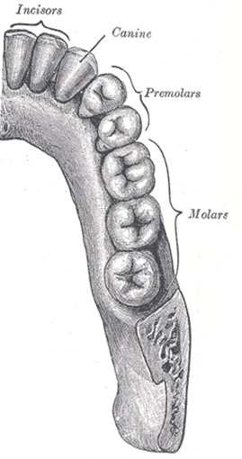
Teeth (figure 3) are responsible for biting, slicing, tearing, chopping and grinding food. The objective is to create smaller pieces to swallow. This process aids digestion by creating a greater surface area for digestive enzymes to work. It is not necessary to thoroughly chew your food, but it makes it easier for your digestive system to process your food. Of course, chewing and paying attention to the food in the mouth will reduce the incidence of choking.
Figure 3: Teeth types

The incisors slice chunks of food.
Canines rip and tear food.
Pre–molars and molars are responsible for chewing and grinding food to a pulp.
Lips, cheeks and tongue work together to push the mass of food (called a bolus) back toward the molars.
The evolution of mammals has created many different "eaters". If you peer (carefully) inside a domestic cat's (a carnivore) mouth, you will see many sharp teeth. These animals do not chew their food. In contrast, the mouth of a deer (an herbivore) has some incisors at the front of the mouth, but the rest of the mouth is full of grinding molars. Humans have both sharp and grinding teeth because they are omnivores.
A classroom activity to help students understand the evolution of mammals is to make observations of different skulls. This can be done by purchasing skull casts, borrowing skulls, or visiting natural history museums such as the Peabody Museum of Natural History in New Haven or the American Museum of Natural History.
Smell, sight, and even the thought of food causes salivary glands to activate. Salivary glands are located at the back of the mouth, under the tongue, on the face, and under the sides of the lower jaw. These are connected to the mouth by tubes called "ducts".
The largest is the parotid gland, which is near the cheek bones. The submandibular salivary gland has a duct that opens at the base of the tongue. The sublingual gland is on floor of mouth, under the tongue (figure 4).
Dogs and cats have no amylase in their saliva. Their natural diet contains almost no carbohydrates
4
.
Human have six to eight times the level of salivary amylase than chimpanzees. This is most likely a result of our different diets. Relative to humans, chimps eat very little starch and much more fruit
5
.
The Digestive system
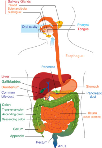
The digestive system can be thought of as a long tube, called the digestive tract or gut, that begins in mouth (figure 4). This opening then becomes the pharynx, esophagus, stomach, small and large intestine and the undigested material exits through the anus. This is a 20 to 40 hour process.
As food travels through the digestive system, it is broken down into smaller molecules. The body uses these molecules for generating energy or building structures.
Esophagus
Figure 4: The Digestive System

The pharynx is the structure that connects the mouth to the esophagus. The term esophagus comes from the Greek "entrance for eating". Once the tongue presses the food to the throat and it is swallowed, the food takes about 10 seconds to reach the stomach via the esophagus. The rest of the digestive process is involuntary.
The top of the pharynx opens to the back of the nose (the nasopharynx). The soft palate blocks the opening to the nose, keeping food from being forced upward. The epiglottis is a cartilaginous flap that temporarily blocks the larynx. The vocal cords also close to prevent food from entering the trachea and lungs. The trachea is commonly called the windpipe and leads to the lungs. When attempting to simultaneously eat and talk, choking can result. This is a result of food going down the trachea when the epiglottis is open.
The esophagus is composed of layers of smooth (involuntary) and striated (voluntary) muscle tissue. Waves of muscle contraction and relaxation move the food bolus along. These alternating waves, called peristalsis, continue throughout the digestive system. Peristalsis can be thought of like squeezing a tube of toothpaste. A classroom activity to simulate peristalsis is squeezing a tennis ball through pantyhose. In our seminar we found this to be most effective if segments of pantyhose were cut to the approximate length of a human esophagus. Students are then challenged to move the "bolus" in ten seconds from one end to another!
Cows and other ruminants are said to have four "stomachs". It appears that the first three of these chambers have evolved as outpockets of the esophagus. When food enters these chambers it is acted upon by microbes. As the material is regurgitated and swallowed, it can again be acted upon by the microbes. This process allows ruminants access to the energy contained in cellulose molecules. This energy source is not available to other mammals who do not have this extended digestion time and the appropriate flora and enzymes. P. 46 Rogers.
Stomach
The stomach is is a "J shaped" organ located up behind the left hand side of ribcage. When empty, it is about size of a fist; it can hold up to four liters (the size of a boxing glove) when full. It is lined with folds called rugae. These folds flatten out and disappear as the stomach fills with food.
Gastric acid is produced in epithelial cells and gastric pits in the stomach wall. The gastric glands of the stomach also produce hydrochloric acid that denatures proteins and further softens food while also killing bacteria and viruses. The stomach itself does not dissolve from the action of the hydrochloric acid because it is lined with a lipid–rich mucous that resists the corrosive acid. The cells that line the stomach are replaced frequently, every 3 or 4 days.
Mucous is a slippery glycoprotein, a molecule containing sugar units and protein that retains water. It also resists the proteolytic enzymes of the digestive system while facilitating the movement of food through the digestive system.
The stomach generates about 1.5 liters of gastric juice per day. Gastric juice is watery mucous, digestive enzymes and acid. Zymogenic cells in the stomach release pepsinogen, which is an inactive form of the protelytic enzyme pepsin. Pepsinogen is activated by stomach acid to become pepsin. Pepsin initates the breakdown of protein by breaking polypeptides (proteins) down into peptides (strings of amino acids). When amino acids are dissolved in acidic solutions, they disassociate because one end is positive (the nitrogen terminal, H
3
N
+
), one is negative (the carboxyl terminal is COO
–
). Water molecules are also polar, so some amino acids are attracted to the negative and positive ends of water.
Three concentric sets of muscles make up the stomach wall. Three times per minute the stomach vigorously squeezes and tightens around a creamy liquid of food, mucous, and enzymes called chyme. As the stomach mixes and works the chyme, water quickly passes to the small intestine. Few food nutrients are absorbed in stomach.
Starchy meals pass through the stomach in about an hour. Lipid meals (meats, cheese, fried food) stay in stomach for three or more hours. When the chyme is soupy enough, peristaltic waves take the digesting food to the end of the stomach. At the distal end, the muscles around the pyloric sphinctor muscles relax, and chyme is released in peristaltic waves into the small intestine.
In the stomach, the proteolytic enzyme pepsin cleaves between these amino acids.
Phenylanine and Alanine
Phenylanine and Leucine
Phenylanine and Methionine
Tyrosine and Alanine
Tyrosine and Leucine
Tyrosine and Methionine
Tryptophan and Alanine
Tryptophan and Leucine
Tryptophan and Methionine
A classroom activity can be used to illustrate this enzymatic cleaving. Dr. Stewart shared a paper digestion activity he created. To build upon his activity, I add decorative scissors with cuts such as "Pinkering", "Flash", "Colonial", "Victorian", "Arabian", and "Double Bubble" to indicate different enzymes acting on different substrates. Additional teaching strategies involve using lactase enzyme on various "milks" such as soy, rice and cow. Also suggested is a laboratory integrating "Beano" enzyme to a variety of foods and recording the effectiveness of the enzyme
6
.
Harmful bacteria or viruses can cause an upset stomach. Heartburn and indigestion can be caused by eating too quickly.
Small Intestine
Called "small" because at 1.5 inches, it's about half as wide as the large intestine. It is folded and rolled up in an area just below your rib cage in the middle of your body. At twenty feet, the small intestine is the longest and most important part of the digestive system.
The small intestine is where most of our digestion occurs. The inner lining is ridged, folded, and studded with millions of little projections called villi and microvilli. If the small intestine were flattened out, it would have an area of almost 3000 square feet! This represents an area ten times greater than the area of your skin and is about as big as a basketball court!
It takes a while to absorb food through the gut wall. The small intestine has increased the surface area by increasing its length. The small intestine fits because it is curled up and folded over itself.
The small intestine allows small molecules to pass through the wall, but we eat big molecules. A metaphor for this organ is a chain gang – which makes little rocks out of bigger rocks.
Protein molecules need to be disassembled into single amino acid or di– or tripeptide in order to be absorbed. Pancreatic proteases break polypeptides into peptides (amino acid chains). Peptidases take these chains down to individual amino acids. Proteases trypsin and chymotrypsin reduce proteins to their constituent amino acids.
Carbohydrate enzymes that are active in breaking down these large molecules in the small intestine include maltase. These enzymes act on maltose to reduce them to the monosaccharide glucose.
Students can be engaged in this activity by using the decorative scissors as an analogue for the protease trypsin to cleave the peptides arginine and leucine at the carboxyl (right) end. The same can be done with a different pattern cut to indicate the action of protease chymotrypsin for tyrosine and tryptophan.
Fatty acids and monoglycerides (after pancreatic enzyme digestion) are passed to lacteal (lymph) capillary and sent to the circulatory system and eventually the liver.
Villi have many capillaries to facilitate transport of digested proteins and sugars into the bloodstream. Lacteals are tubes in the villi that allow fats to be transported to the blood. Villi bend and wave to keep blood flowing through the capillaries and into the bloodstream. Ateries and veins are located just under the base of the villi.
The lining of small intestine make intestinal juice that includes enzymes to digest proteins, carbohydrates, and fats. Mucous is also produced, and as with the mouth, esophagus and stomach, it coats and protects the lining of small intestine.
The first section of the small intestine is called the duodenum. The duodenum is about a foot long and shaped like a letter "C". The liver and pancreas have ducts that join together and have a common opening to the duodenum.
The second section of the small intestine is called the jejunum and is about 6.5 feet long. Most digestion happens in these two sections.
The third and final section of the small intestine is called the ileum. By the end of the small intestine, water and waste are all that remain to enter the large intestine.