Twenty-four hours a day the brain is busy sending and receiving messages. These messages travel along a network of nerve cells that make up the nervous system (Freidman, 1990). The human brain is composed of billions of cells. Estimates suggest that there are between 100 billion and one trillion neurons in a single human brain. It sends and receives an immense number of messages every day along neurons. (Dupont, 1997). "A nerve cell, or neuron, is a cell that receives information from other nerve cells or from the sensory organs and then projects that information to other nerve cells, while still other neurons project it back to the parts of the body that interact with the environment, such as the muscles." (Bransford, Brown, and Cocking, 1999). Neurons are designed to receive, examine and send information to other neurons. The neuron is the fundamental building block of the nervous tissue in the brain and consists of three major parts: the cell body, the axon and the dendrites. The cell body houses genetic information in its nucleus. The axon extends from the neuron to make contact with other neurons and send messages. The dendrites are the neuron extensions that form a tree-like structure which is the input side of the neuron that receive messages from other nerve cells. Most of the excitatory information is passed into the cell through projections on the dendrites called spines. These neurons send messages to each other over a very small space between the axon and dendrite. This space between the neurons or gap is known as a synapse (Dupont, 1997). Synapses can be excitatory or inhibitory. The neuron then integrates the received information from the synapse and the output is determined.
Special chemicals send messages across these gaps. The chemicals, called transmitters, take messages from the axon of one nerve cell and send it to the receptors of another nerve cell. When the transmitters bind with the receptor the message is communicated. A cell may receive thousands of such messages in just a few seconds. For example, your eyes are composed of millions of nerve cells, yet the eyes don't really see pictures. They simply send electrical signals to the brain. The brain then receives the information from the nerve cells, examines it, and compares it to other information it already has, decides whether or not it's important enough to store in its memory and then uses the information to help us react to the outside world. This is one aspect of learning.
At birth the brain has only a small proportion of the trillions of synapses it will have. The rest of the synapses are formed after birth. A portion of this is guided through experiences. Synaptic connections are added in two ways. Some synapses are overproduced and then selectively lost. The brain does this in order to incorporate information from experience, usually during the early periods of development. Another method is through the addition of new synapses. Synapse addition continues throughout life and is very important in later life. This process is driven by experience. After the cycle of synapse overproduction and selection runs its course, additional changes occur in the brain. This includes the modification of existing synapses and the addition of new synapses. Activity in the nervous system associated with learning experiences somehow causes nerve cells to create new synapses. This is unlike the process of synapse overproduction and loss. Synapse addition and modification are lifelong processes and they are driven by experience. "In essence, the quality of information to which one is exposed and the amount of information one acquires is reflected throughout one's life in the structure of the brain. This process is probably not the only way that information is stored in the brain, but it is a very important way that provides insight into how people learn" ( Bransford…et all ,1999).
The neurons can not do this alone. They need a support group. The structures inside the brain are made up of about 100 billion neurons and trillions of support cells known as glia. Although the neurons are responsible for sending the messages they couldn't do it without the help of the glia cells. There are different types of glia cells that optimize brain function. The oligodendrocytes speed up the electrical signal that is traveling down the axon. Microglias are immune cells that detect damaged neurons and infections. The astrocytes keep the neurons in place, and assist in the transport of nutrients. They also communicate and modify signals between the neurons. Each astrocyte can interact with several neurons and thousands of synapses to properly integrate information (National Institute of Health, 2009).
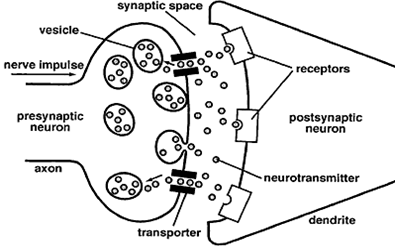
Figure 3: "Schematic diagram of a synapse. In response to an electrical impulse, neuro-transmitter molecules released from the presynaptic axon terminal bind to the specific receptors for that neurotransmitter on the postsynaptic neuron." Courtesy of National Institute of Health.
Lesson 5 - Build a Neuron
Objective: The students will understand the structure of a neuron by creating a model of one that can later be used to demonstrate a synapse.
Materials: 5 different colors of pipe cleaners: one color each for the dendrites, cell body, axon, myelin sheath and synaptic terminal. (1 set per student) scissors
Introduction: Show students the diagram of a neuron. Point out the main structures: axons, dendrites, cell body and myelin sheath. Emphasize the unique shape of the nerve cell - long and thin. Ask students what the main job of a nerve cell is. (Receiving and transmitting messages.) What do we use in everyday life to communicate with people across town or across the country? (Telephone, People and Computers) How is the signal sent? (Over a long, thin wire.). Review that nerve cells do not actually touch one another, but that there are tiny gaps between nerve cells called synapses. Neurons produce powerful chemicals that are released into the synapse as a message is being sent from one cell to the next. The neurotransmitter is released by an axon. It crosses the synapse and attaches to a receptor on the other side. Dendrites sprout to make connections with other neurons. This is one of several mechanisms involved in learning.
Procedure:
-
1. Take one pipe cleaner and roll it into a ball. This is will be the cell body.
-
2. Take another pipe cleaner and attach it to the new "cell body" by pushing it through the ball so there are two halves sticking out. Take the two halves and twist them together into a single extension. This will be the axon.
-
3. Take other pipe cleaners and push them through the "cell body" on the side opposite the axon. These are dendrites. These can be shorter than your axon and you can twist more pipe cleaners to make more dendrites
-
4. Wrap small individual pipe cleaners along the length of the axon. These will represent the myelin sheath.
-
5. Wrap another pipe cleaner on the end of the axon. This will be the synaptic terminal (Chudler, 2009).
Closure: The students can share their creations with the class all the while explaining the parts of a neuron and how they communicate with each other. For further demonstration of a synapse check out this website and watch the animation with the class. It explains how neurons communicate as well as gives an animated visual to ensure comprehension. http://learn.genetics.utah.edu/content/addiction/reward/neurontalk.html